[2018]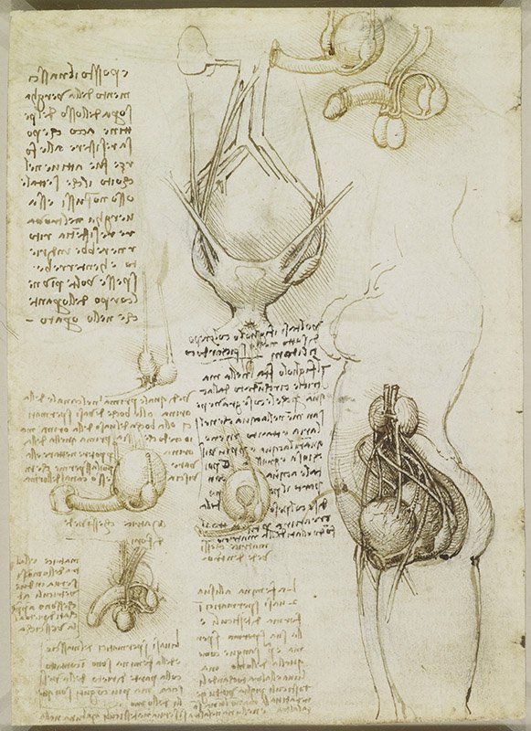


- Da Vinci, Leonardo. c. 1508. The Male and Female Reproductive Systems. Pen and ink over black chalk, 19.1 × 13.8 cm. The Royal Collection HM Queen Elizabeth II. RL 19095v; QA III.1v; O’M&S 201; K&P 54v.
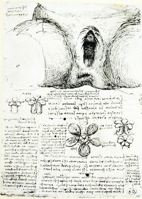
- Da Vinci, Leonardo. c. 1508. The Vulva and Anus. Pen and ink over black chalk, 19.1 × 13.8 cm. The Royal Collection HM Queen Elizabeth II. RL 19095r; QA III.1r; O’M&S 200; K&P 54r
- Casserius, Giulio. c. 1600. Penis and Dissection of the Perineum Origin. De formato foetu liber singularis. Volume II. The United States National Library of Medicine. https://collections.nlm.nih.gov/catalog/nlm:nlmuid-2417033R-bk
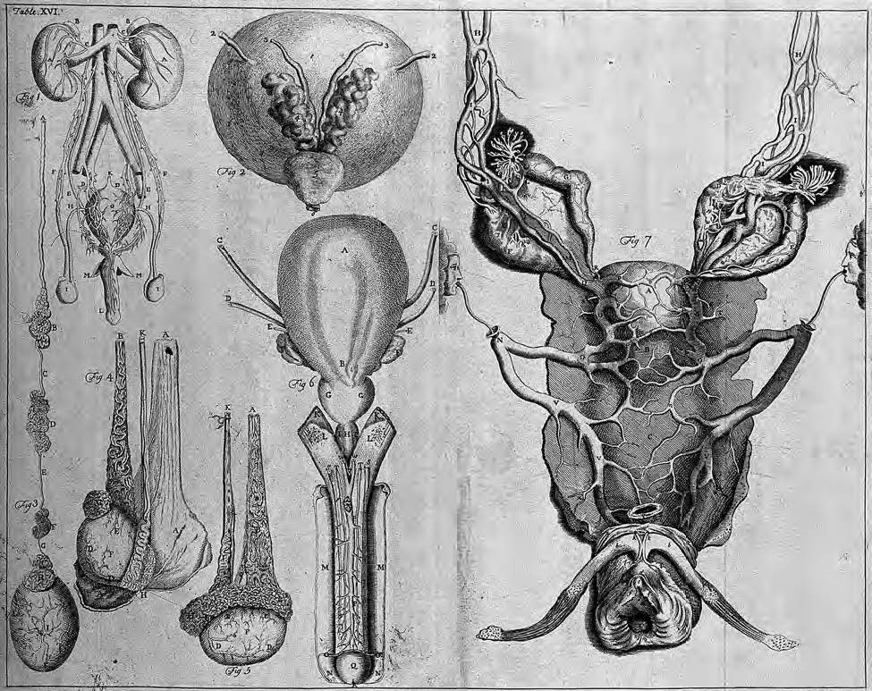
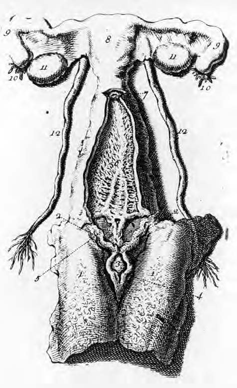
- De Graaf, Regnier. c. 1743. The Genito-urinary System. etching ; 30.2 x 38.3 cm. https://wellcomecollection.org/works/qe4jyhj8/items
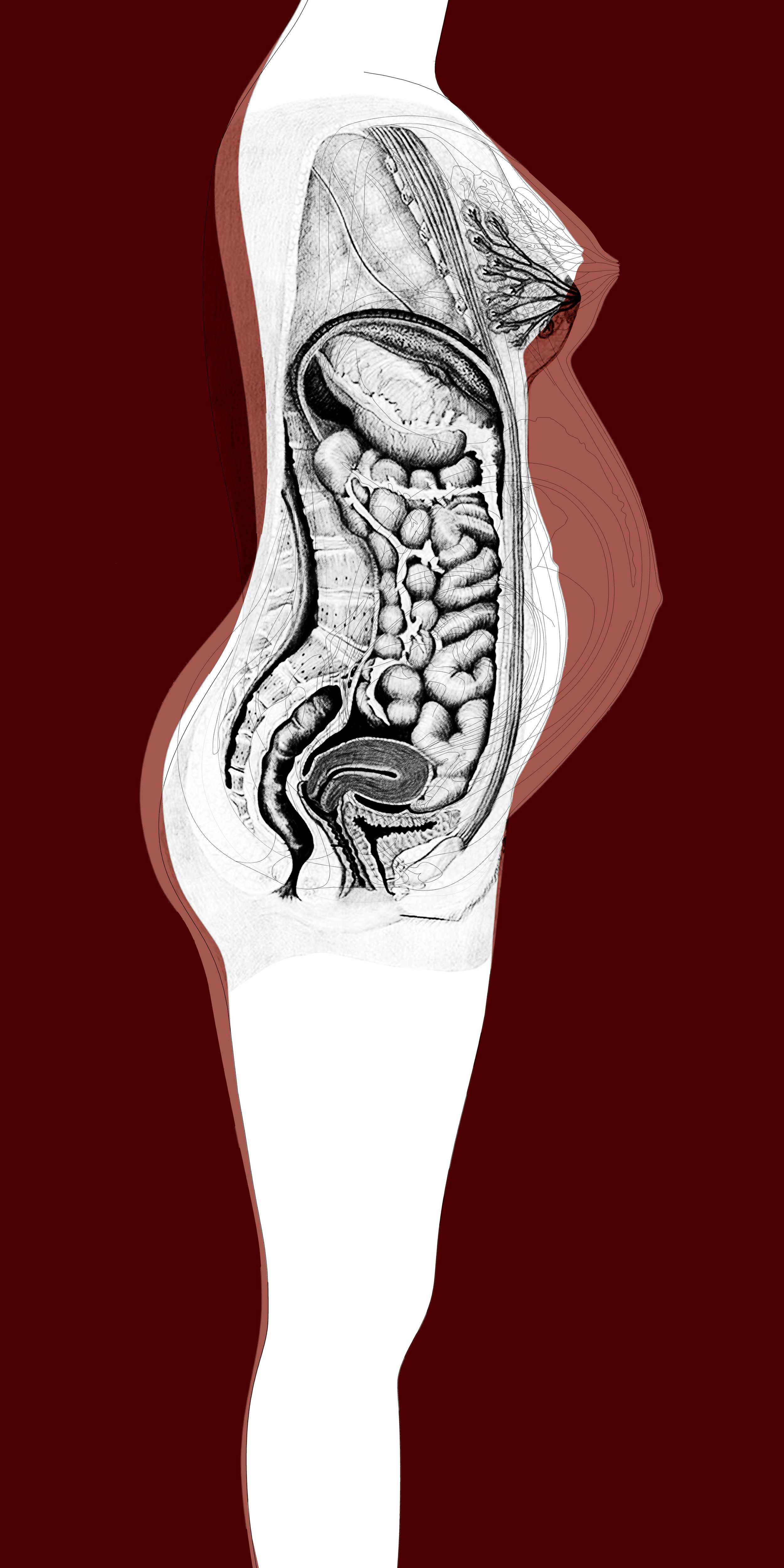
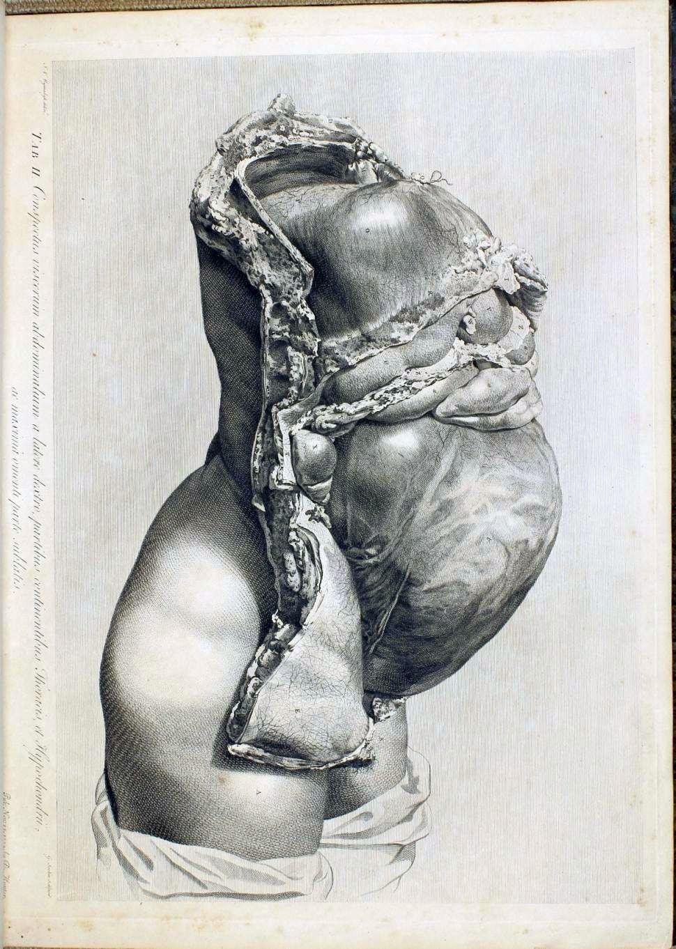
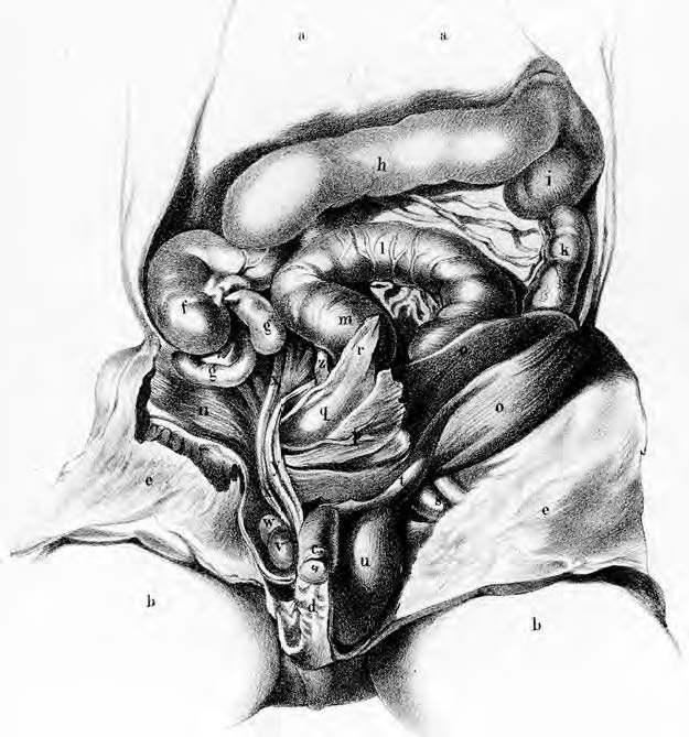
- Hunter, William. c. 1774. View of the Abdominal Viscera From the Right Side. Anatomia Uteri Humani Gravida Tabulis Illustrata. The United States National Library of Medicine. https://collections.nlm.nih.gov/catalog/nlm:nlmuid-2491060R-bk
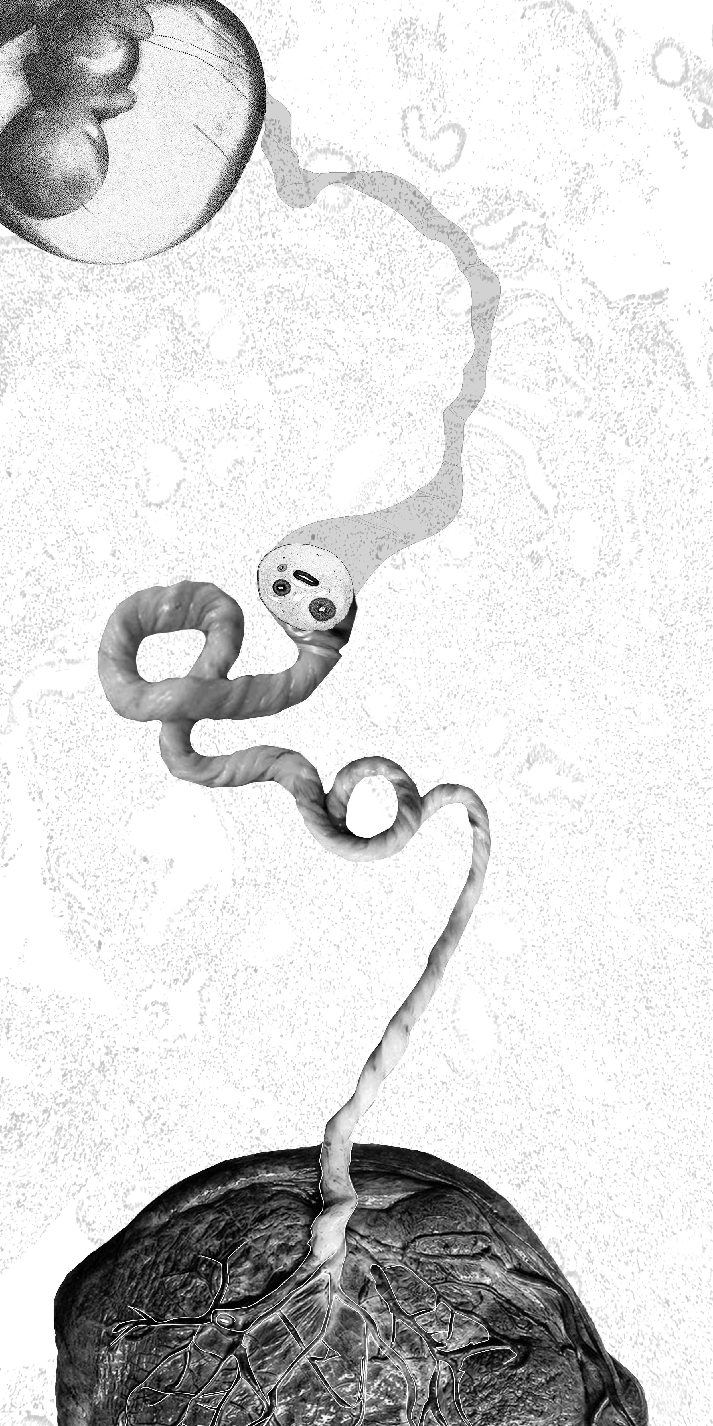
- Hunter, William. c. 1774. Placenta and Fetus. Anatomia Uteri Humani Gravidi Tabulis Illustrata. The United States National Library of Medicine. https://collections.nlm.nih.gov/catalog/nlm:nlmuid-2491060R-bk
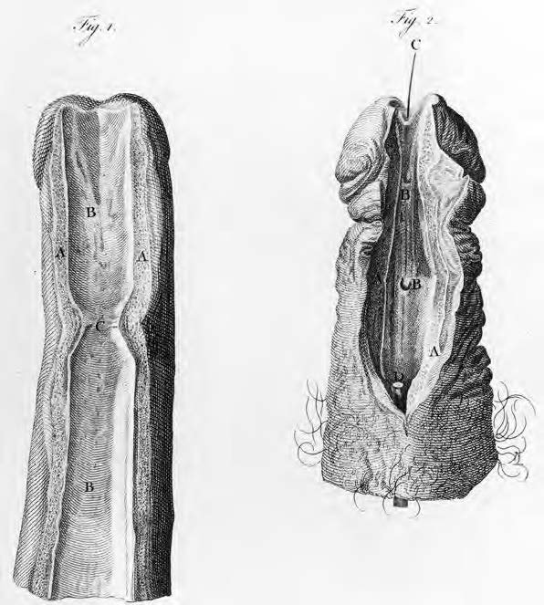
- Hunter, John. c. 1774. The Penis Flit Open. A Treatise on the Venereal Disease. The United States National Library of Medicine. https://collections.nlm.nih.gov/catalog/nlm:nlmuid-2558019R-bk
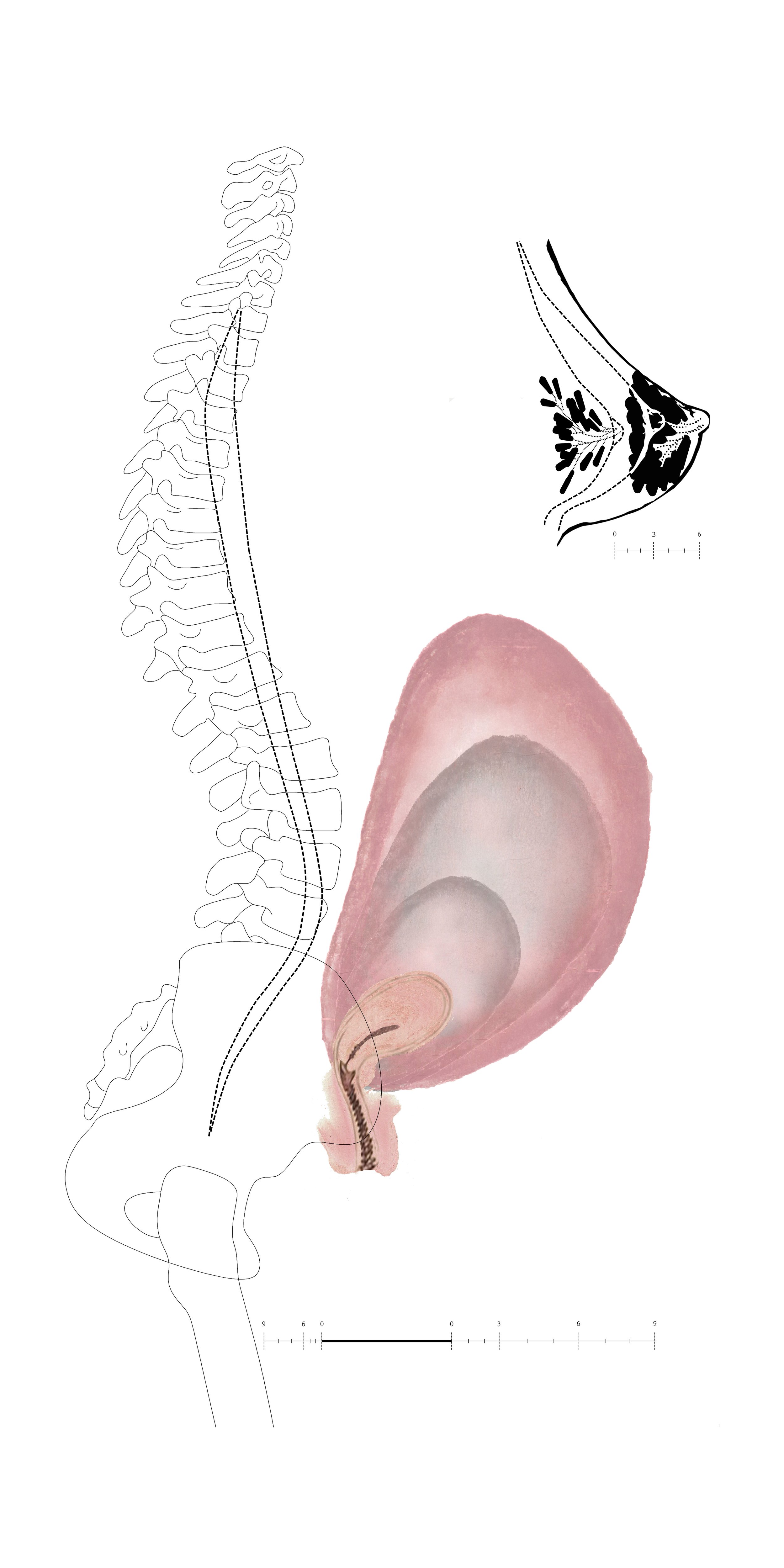
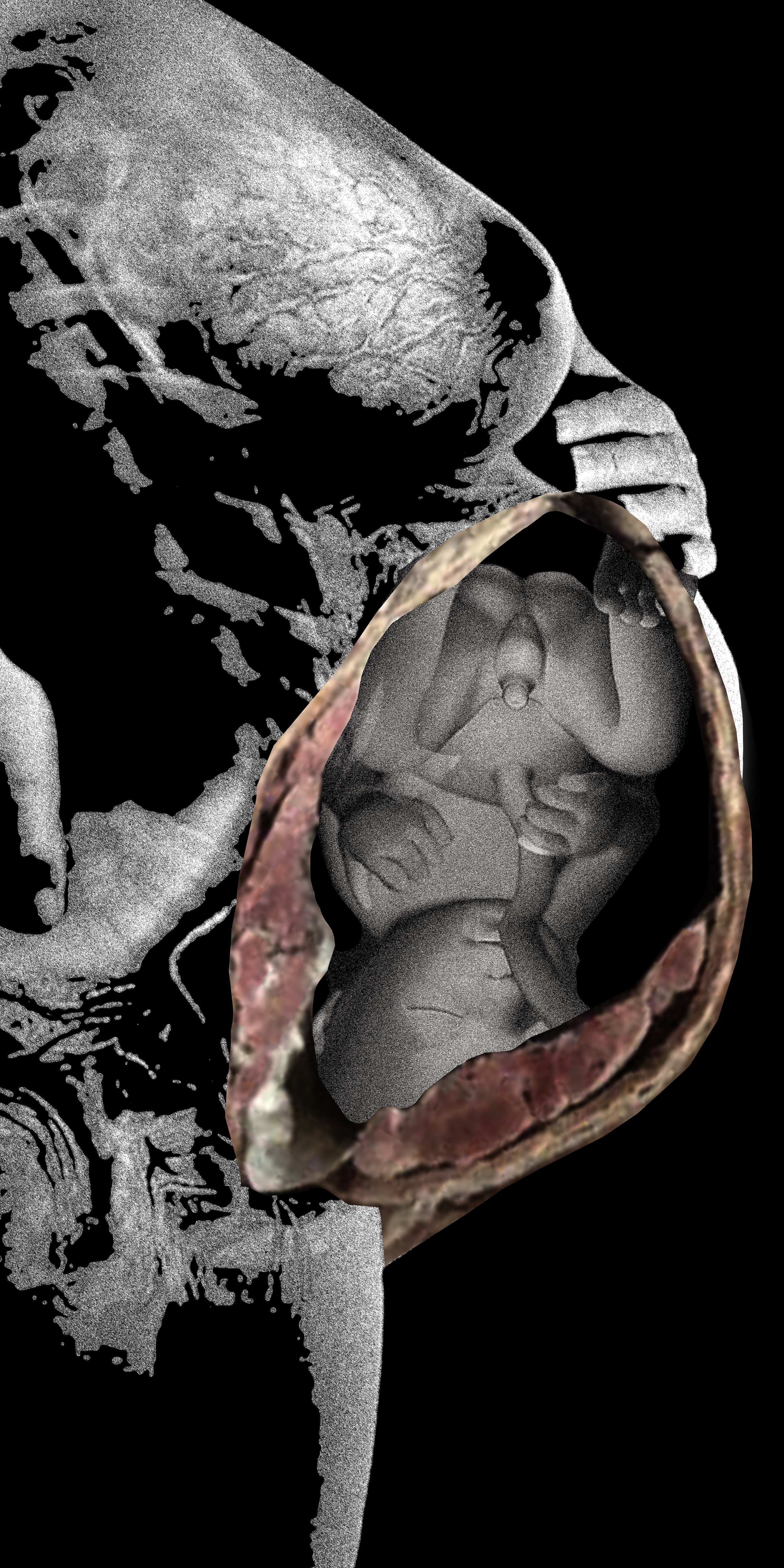
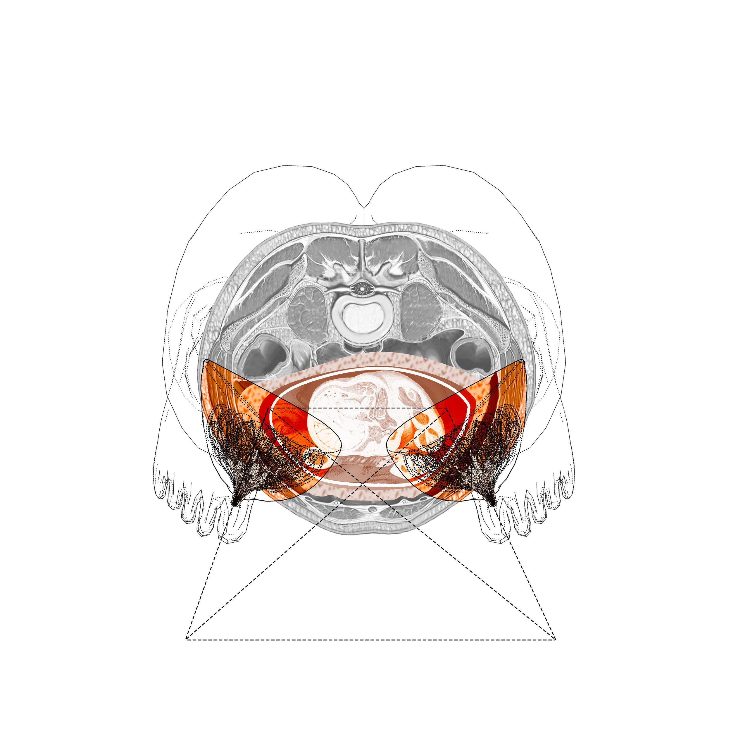
In the article “This Sex Which Is Not One,” Luce Irigaray argues that the conceptualization of female sexuality is based on masculine parameters. This position applies to architectural studies as well. Historically, the female body has been neglected in human anatomy studies and, consequently, in the architectural standards established upon the human body. This project focuses on the female body as it goes through the transformation in the reproduction process that her body is equipped for. This transformation is most apparent during pregnancy as the female body goes through both physical and non-physical alteration while a new body develops within hers, prompting the necessary changes to create within the mother’s body the conditions and environment for its development and growth and for the close interaction between these two bodies for nine months. With pregnancy, a temporary vital organ, the placenta, develops in the female body as a mediator between mother and fetus, from which the umbilical cord emerges as the connecting channel between them. This organ makes all transactions concerning oxygen, nutrients, hormones, and most other exchanges in this process possible.
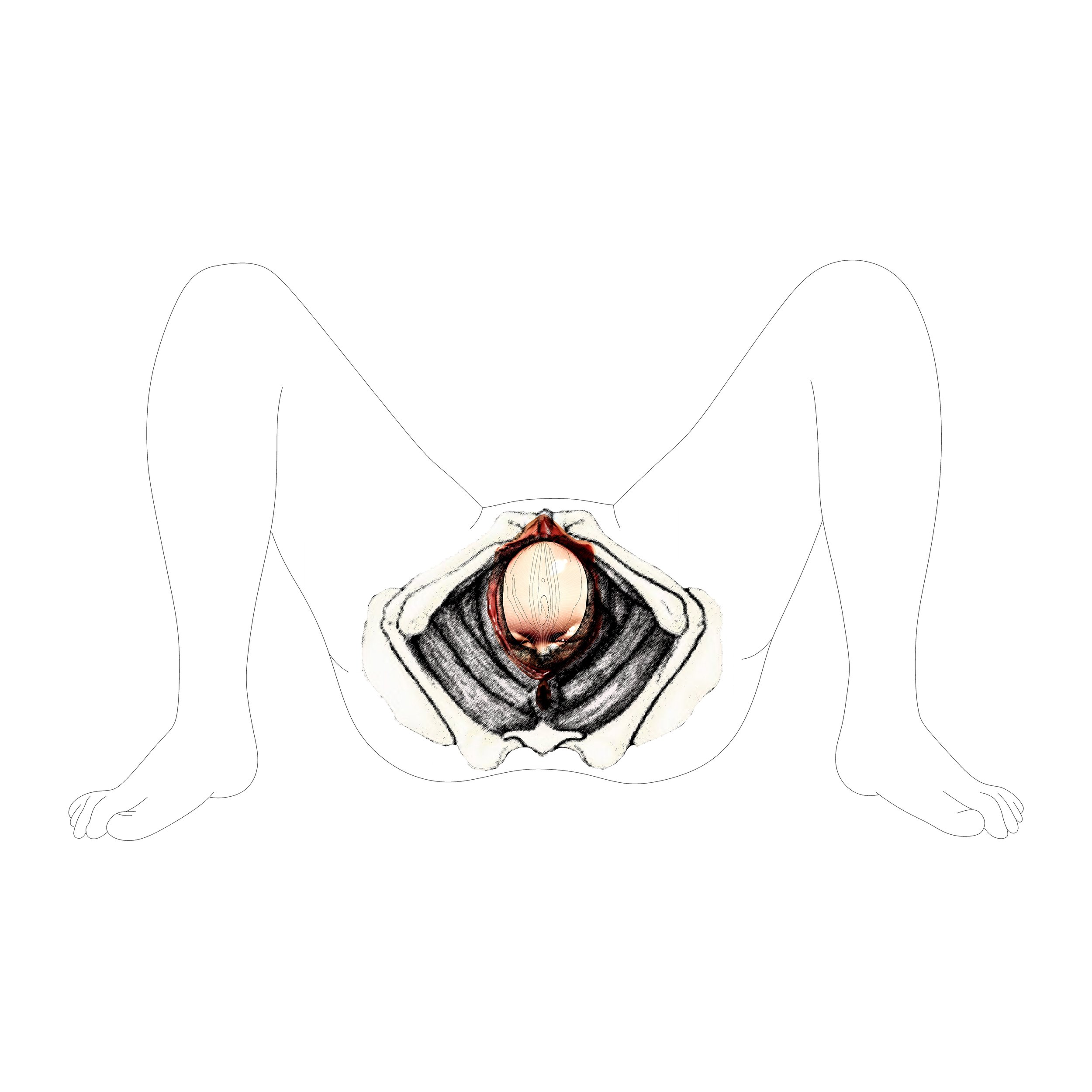
The project attempts to look at some specific moments of this process as an architectural phenomenon: the restructuring of one body and the structuring of another. The moment of birth is the ultimate culmination of the process when the fetus leaves the womb, as a baby enters an environment external to the mother’s body, as the umbilical cord is cut. From the female body, seen as a place of nourishment, another male or female being will be born.
A Body Within the Body flourished through conversations with Professor Diana Agrest and her critiques as part of her Advanced Research-Design Graduate Studio on the human body at The Irwin S. Chanin School of Architecture of The Cooper Union in 2018.

image: Lea Bertucci, photographer. Courtesy of The Irwin S. Chanin School of Architecture Archive of The Cooper Union.